
Key Takeaways
- Fascia forms a connected tissue web that supports muscles nerves joints organs and body shape.
- This tissue helps neighboring layers slide so motion stays smooth efficient and less irritating.
- Fascia also carries sensory nerve endings linked with position sense pressure and pain signaling.
- Research suggests force can spread through fascial links instead of staying inside one muscle.
- Reduced fascial glide is linked with stiffness movement limits and some chronic low back pain.
Fascia As Tissue
Support Web
Fascia is connective tissue that surrounds and links muscles, tendons, bones, nerves, blood vessels and organs. Older anatomy teaching often treated it as wrapping or packing, yet newer reviews describe it as a continuous network with structural and sensory features that deserve direct attention in their own right (1, 2).
Deep fascia contains organized collagen, elastin and ground substance arranged in layers that can resist tension while still allowing controlled deformation. This design helps tissues hold shape, separate from each other and keep moving parts from collapsing into one mass during daily motion, loaded exercise and changes in posture (2).
Sensor Rich Tissue
Fascia is not a silent sheet. A systematic review of fascial innervation found sensory endings linked with proprioception and nociception across muscular and deep fascia, which means this tissue can contribute to body position sense and pain signaling rather than serving only as passive support (3). Histology studies of limb fascia support the same idea by showing nerve elements within deep fascial layers, which helps explain why irritated fascia can feel sore, tender or unusually reactive during movement even when imaging shows no major muscle tear (4, 5).
Motion & Load
Layered Glide
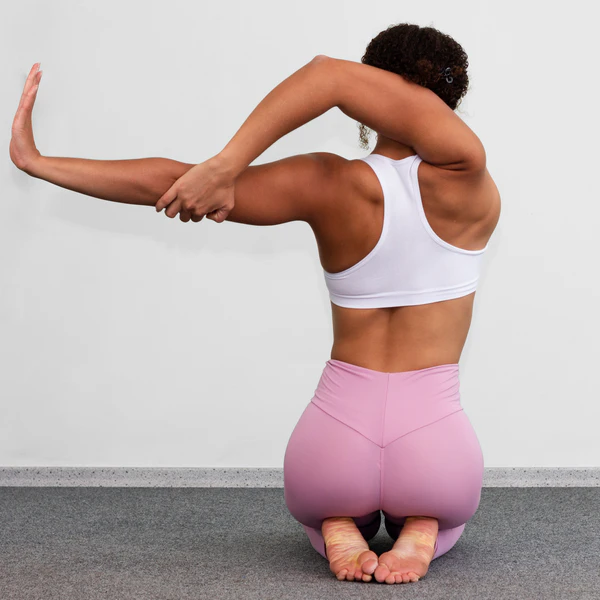
Movement depends on tissues sliding as much as stretching. Fascia helps create shear planes between neighboring muscles and between muscle and skin, so parts can move with each other without dragging or pinching. When those planes move well, bending, reaching and rotation feel easier and less effortful (6).
Research on the thoracolumbar fascia gives a useful example. In one human study, people with chronic low back pain showed lower shear strain in this fascia than people without back pain, suggesting reduced glide may be one part of the stiffness and movement changes seen in persistent symptoms (7). A later systematic review on ultrasound imaging of the thoracolumbar fascia found growing evidence that imaging can capture thickness, displacement, stiffness and shear related features in both healthy and painful states, although methods still vary across studies (8).
Daily movement keeps these layers exchanging load from one region to another. Long periods of immobility, repetitive low variety motion and guarding after pain can all reduce normal sliding, leaving motion feeling sticky rather than free. Research has not reduced pain to fascia alone, but current evidence supports fascial mobility as one piece of the larger musculoskeletal picture (6, 8).
Force Across Regions
Muscles do not work in isolation, and fascia helps explain why. Reviews on myofascial force transmission report evidence that force can spread across connective tissue links between neighboring muscles and along broader chains, so movement at one joint may influence tissue displacement well above or below that area (9, 10).
Human imaging studies add direct support. Passive ankle motion has been associated with soft tissue displacement in the dorsal thigh, and other work has reported linked displacement between muscle and superficial fascia, both pointing to mechanical continuity across regions rather than isolated local action alone (11, 12). Flexibility tests and exercise programs should be read with this in mind, since a stretch or restriction felt in one place may reflect load sharing through connected tissues elsewhere.
Pain & Sensitivity
Pain Without Injury
Pain is shaped by the nervous system, past experience, local tissue state and the demands placed on the body. Fascia fits into this picture because it is innervated, mechanically active and sensitive to inflammatory change. A fascia that is irritated, compressed or moving poorly can become part of the symptom source even when the problem does not look dramatic on a scan (3, 6).
Low back pain research offers the clearest example at present. Reduced shear strain in the thoracolumbar fascia does not prove a single cause, but it does show a measurable difference linked with chronic symptoms. Current reviews describe fascia as a possible contributor to pain through altered glide, local inflammation, fibrosis and increased sensitivity of small nerve fibers (7, 6).
Biomechanics also helps explain why pain can spread beyond one tender spot. If force is transferred through connected fascial tissues, then overload, guarding or poor movement tolerance in one region may raise strain in another. Reviews of in vivo force transfer suggest this idea is plausible in humans, though the size of the effect likely changes by tissue, task and individual anatomy (10, 9).
Fascia is neither a miracle answer nor a useless wrapper. Evidence supports its contribution to support, sensation, load sharing and some pain states, while many questions about cause, timing and treatment response remain open. Strong claims that every ache comes from fascia go beyond what current human research can support.
Daily Care
Better Motion Inputs
Fascia responds to repeated inputs from movement, loading and rest. Regular walking, strength work through comfortable ranges, varied joint motion and gradual return to activity after pain are all consistent with keeping tissues mobile and load tolerant. Sudden aggressive stretching is not required, and more force is not always better. A steadier mix of motion and recovery gives fascial layers more chances to slide, adapt and share load well across the body.
For any health concerns or questions about a medical condition, get guidance from a physician or another appropriately trained clinician. Before changing your diet, supplements or health routine, talk with a licensed healthcare professional.
FAQs
Is fascia the same as muscle?
No. Muscle creates active force, while fascia is connective tissue that surrounds, links and separates structures throughout the body. Both work together during movement.
Can fascia cause pain on its own?
Fascia can be part of a pain source because it contains sensory nerve endings and can become irritated or less mobile. Pain still has many drivers, so fascia is one part of a larger picture.
Does stretching fix fascia problems?
Stretching may help some people, but it is not the only option. Gentle movement variety, strength training, walking and graded return to activity often support better tissue tolerance too.
Why does pain show up away from the tight area?
Connected tissues can share load across regions, so stress in one place may change tension or movement somewhere else. Pain location does not always identify the full source.
Can imaging show fascia changes?
Ultrasound can assess some fascial features such as thickness, displacement and shear related behavior. Findings still need to be interpreted with symptoms and movement, not in isolation.
Research
Benjamin, M. (2009) The fascia of the limbs and back a review. Journal of Anatomy, 214(1), pp. 1 to 18. Available at https://pubmed.ncbi.nlm.nih.gov/19166469/
Willard, F.H. et al. (2012) The thoracolumbar fascia anatomy, function and clinical considerations. Journal of Anatomy, 221(6), pp. 507 to 536. Available at https://pubmed.ncbi.nlm.nih.gov/22630613/
Suarez Rodriguez, V. et al. (2022) Fascial Innervation A Systematic Review of the Literature. International Journal of Molecular Sciences, 23(10), 5674. Available at https://pubmed.ncbi.nlm.nih.gov/35628484/
Stecco, C. et al. (2007) Anatomy of the deep fascia of the upper limb. Second part study of innervation. Morphologie, 91(292), pp. 38 to 43. Available at https://pubmed.ncbi.nlm.nih.gov/17574469/
Stecco, C. et al. (2008) Histological study of the deep fasciae of the limbs. Journal of Bodywork and Movement Therapies, 12(3), pp. 225 to 230. Available at https://pubmed.ncbi.nlm.nih.gov/19083678/
Langevin, H.M. (2021) Fascia Mobility, Proprioception, and Myofascial Pain. Life, 11(7), 668. Available at https://pubmed.ncbi.nlm.nih.gov/34357040/
Langevin, H.M. et al. (2011) Reduced thoracolumbar fascia shear strain in human chronic low back pain. BMC Musculoskeletal Disorders, 12, 203. Available at https://pubmed.ncbi.nlm.nih.gov/21929806/
Pirri, C. et al. (2024) Ultrasound Imaging of Thoracolumbar Fascia A Systematic Review. Medicina, 60(7), 1090. Available at https://pubmed.ncbi.nlm.nih.gov/39064519/
Krause, F. et al. (2016) Intermuscular force transmission along myofascial chains a systematic review. Journal of Anatomy, 228(6), pp. 910 to 918. Available at https://pubmed.ncbi.nlm.nih.gov/27001027/
Ajimsha, M.S. et al. (2022) Evidence of in vivo myofascial force transfer in humans a systematic scoping review. Journal of Bodywork and Movement Therapies, 32, pp. 183 to 195. Available at https://pubmed.ncbi.nlm.nih.gov/36180147/
Wilke, J. et al. (2020) Ankle Motion Is Associated With Soft Tissue Displacement in the Dorsal Thigh An in vivo Investigation Suggesting Myofascial Force Transmission Across the Knee Joint. Frontiers in Physiology, 11, 180. Available at https://pubmed.ncbi.nlm.nih.gov/32210836/
Wilke, J. and Tenberg, S. (2020) Semimembranosus muscle displacement is associated with movement of the superficial fascia An in vivo ultrasound investigation. Journal of Anatomy, 237(6), pp. 1026 to 1031. Available at https://pubmed.ncbi.nlm.nih.gov/32794194/
Fede, C. et al. (2020) Fascia and soft tissues innervation in the human hip and their possible role in post surgical pain. Journal of Orthopaedic Research, 38(7), pp. 1646 to 1654. Available at https://pubmed.ncbi.nlm.nih.gov/32181900/
do Carmo Carvalhais, V.O. et al. (2013) Myofascial force transmission between the latissimus dorsi and gluteus maximus muscles an in vivo experiment. Journal of Biomechanics, 46(5), pp. 1003 to 1007. Available at https://pubmed.ncbi.nlm.nih.gov/23394717/
Huijing, P.A. et al. (2011) Effects of knee joint angle on global and local strains within human triceps surae muscle MRI analysis indicating in vivo myofascial force transmission between synergistic muscles. Surgical and Radiologic Anatomy, 33(10), pp. 869 to 879. Available at https://pubmed.ncbi.nlm.nih.gov/21912991/
Stecco, C. et al. (2006) Histological Characteristics of the Deep Fascia of the Upper Limb. Italian Journal of Anatomy and Embryology, 111(2), pp. 105 to 110. Available at https://pubmed.ncbi.nlm.nih.gov/16981399/
Benetazzo, L. et al. (2011) 3D reconstruction of the crural and thoracolumbar fasciae. Surgical and Radiologic Anatomy, 33(10), pp. 855 to 862. Available at https://pubmed.ncbi.nlm.nih.gov/22024628/