Key Takeaways
- Bone health depends on constant remodeling that removes old tissue and replaces it.
- Fracture healing follows staged biology that starts fast but finishes with slow reshaping.
- Osteocytes sense strain in bone and help coordinate repair signals during recovery.
- Cell signals couple breakdown and rebuilding so bone stays strong without unnecessary thickening.
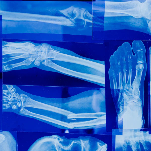
- X rays can look improved before bone regains full strength, so loading needs care.
Bone Growth Pathways
Growth Plates
Long bones grow in length at growth plates, where cartilage is replaced by mineralized bone as children mature.
The process depends on orderly cell turnover, blood supply and the arrival of mineral and collagen building blocks in the right sequence.
Growth slows and then stops when growth plates close, so adult height no longer increases. Bone still stays active afterward because remodeling continues for life. (1)
Bone also grows in thickness, and this part never fully stops.
Outer bone surfaces can add tissue when the skeleton faces new mechanical demands, while inner surfaces can be resorbed to keep bones light enough for movement.
This balance helps bones stay mechanically competent without becoming heavy. Researchers describe this as an operational process that supports survival and mechanical function. (2)
Thickening Over Time
Mechanical signals shape how bone adapts, because bone responds to strain from walking, lifting and impact.
Osteocytes sit inside mineralized bone and can detect fluid movement and deformation, then send signals that shift the balance between bone resorption and bone formation.
Reviews of mechanical regulation describe how loading can encourage formation while unloading tends to promote resorption. (3, 4)
Fracture Repair Stages
Early Response
A fracture starts with bleeding and clot formation around the break. Immune cells move in to clear damaged tissue and reduce infection risk.
This early inflammation is normal and usually short lived, but it sets the stage for later rebuilding. Repair depends on adequate blood flow because cells and nutrients must reach the site. (5)
Osteoclast activity also increases near injured bone, because broken edges and damaged microstructures need to be removed.
Resorption creates a cleaner surface for new tissue to attach and it helps direct the shape of later repair.
Bone turnover is not only a demolition process, since breakdown is linked to rebuilding through coupling signals. (1)
Osteocytes can be injured by the fracture itself, and osteocyte networks can also signal for targeted remodeling around microdamage.
Several reviews describe osteocytes as key coordinators because they connect mechanical sensing with chemical messaging that influences both osteoclasts and osteoblast lineage cells. (4, 6)
Callus Formation
Soon after injury, the body forms a soft callus that stabilizes the break. The callus contains a mix of cartilage like tissue and early bone matrix, and it helps bridge the fracture gap.
Over time, the soft callus is replaced by a harder mineralized callus, which is stronger but still not identical to original bone.
The pace and quality of callus formation vary with injury severity and blood supply. (5)
Osteoblast lineage cells lay down collagen rich matrix and then mineralize it.
Bone formation and resorption stay linked even during callus building, because removal of poorly organized tissue can enable better organized tissue to replace it.
Human studies of remodeling units show resorption and formation unfolding together across space and time rather than as isolated steps. (7)
Remodeling Phase
Remodeling turns the bulky callus into bone that better matches the original shape and internal structure. This stage can take months, and in some fractures it can take longer.
Classic frameworks describe remodeling as targeted or non targeted turnover carried out by multicellular units that move through bone over time.
These ideas help explain why a fracture can feel better while internal structure is still improving. (8)
Remodeling & Signaling
Osteoclast Resorption
Osteoclasts remove mineralized bone by creating a sealed resorption space at the bone surface.
This step is necessary for normal turnover and for repairing microcracks that would otherwise accumulate.
Excess resorption without matching formation can lower bone mass over time, but normal resorption is part of healthy renewal.
Disorders of remodeling show how shifts in turnover balance can change strength even when bone size looks similar. (9)
Many signals regulate osteoclast activity, including the RANKL system and its decoy receptor OPG.
Research in early after menopause physiology has linked increased RANKL activity with higher bone resorption.
Clinical studies have also tested OPG and later agents that act on this pathway, which can reduce fracture risk in selected groups.
These findings show strong biological control points, even though treatment decisions are individualized. (10, 11)
Coupling signals connect osteoclast work to later osteoblast formation, so breakdown can stimulate rebuilding in the right place.
Reviews describe multiple mechanisms for this coupling, including factors released from bone matrix during resorption and signals produced by osteoclasts themselves.
This coordination helps explain why normal remodeling maintains overall structure even with constant turnover. (1)
Osteoblast Formation
Osteoblast lineage cells produce new bone matrix and guide mineral deposition. Formation follows resorption in many remodeling events, but the timing can differ across sites and ages.
The coupling process is a major reason bone can stay stable over long periods even while tissue is replaced. Real time observations of human remodeling units support this linked view of turnover. (7)
Osteocyte Control
Osteocytes are former osteoblasts embedded in bone, connected through a network of cell processes that communicate across large distances.
Osteocytes regulate bone turnover partly through signals such as sclerostin and through their influence on osteoclast and osteoblast behavior.
Reviews describe osteocytes as mechanosensors and as endocrine like cells that can affect whole body mineral handling. (4, 12)
Mechanotransduction research focuses on how osteocytes convert mechanical strain into biochemical signals.
Systematic reviews summarize molecular pathways involved in this conversion, and they highlight the complexity of cell level signaling inside hard mineralized tissue.
This complexity is one reason simple single nutrient explanations for bone health often fall short. (13, 2)
Recovery Factors & Red Flags
Mechanical Loading
Bone responds to load, but the right load depends on healing stage.
Early stability supports callus formation, while gradual weight bearing and progressive strength work can encourage remodeling as pain and medical guidance allow.
Mechanical regulation reviews describe how loading and unloading change remodeling signals, which supports gradual return to activity rather than sudden overload.
Imaging can lag behind strength gains, and strength can lag behind symptom relief, so staged progression is sensible. (3)
Nutrition & Lifestyle
Healing requires sufficient energy and protein because bone matrix is built from collagen and other proteins, and repair also demands immune activity and tissue growth.
Systemic regulation reviews describe how hormones and growth factors influence remodeling and repair, which connects sleep, stress physiology and overall health to recovery biology.
A clinician will often focus on the underlying driver of the fracture, such as low bone density, falls risk or overuse, alongside the fracture itself. (5)
Vitamin and mineral status can influence remodeling, but evidence is strongest for the idea that bone is regulated by interacting systems rather than by a single nutrient.
Bone research reviews emphasize bone as a dynamic organ where resorption and formation are coordinated under mechanical and systemic control.
Fortification and supplementation discussions often reduce this complexity to one lab number, which can miss the broader physiology.
A careful approach looks for overall adequacy and avoids assuming more is always better. (2, 9)
Medications can also affect healing and remodeling, and some act directly on osteoclast pathways.
Large trials of antiresorptive therapy show fracture risk reduction in certain populations, which supports the central role of osteoclast activity in bone outcomes.
Medication choice is not a do it yourself decision, since benefits and risks depend on age, fracture history and other conditions.
People who are healing a fracture should tell their clinician about any bone active drug use. (11)
When Healing Stalls
Delayed union and nonunion have many causes, so warning signs should be evaluated rather than self treated.
Persistent worsening pain, increasing swelling or new deformity can indicate complications.
Fever, drainage or spreading redness raise concern for infection, especially after surgery.
Numbness, color change or loss of pulse symptoms need urgent assessment, because blood flow and nerve function are essential for recovery. (5)
Signs that merit timely medical review include
- Pain that increases week to week instead of gradually settling
- Inability to bear weight after the expected timeline given by a clinician
- New instability or a sensation of bones shifting at the fracture site
- Skin changes such as warmth redness drainage or a wound that will not close
- New tingling weakness or coolness below the injury site
For any health concerns or questions about a medical condition, get guidance from a physician or another appropriately trained clinician. Before changing your diet, supplements or health routine, talk with a licensed healthcare professional.
Suggested Posts


Research
Bolamperti, S., Villa, I. and Rubinacci, A. (2022) Bone remodeling an operational process ensuring survival and bone mechanical competence. Bone Research. Available at https://doi.org/10.1038/
Wang, L. et al. (2022) Mechanical regulation of bone remodeling. Bone Research. Available at https://doi.org/
Robling, A.G. and Bonewald, L.F. (2020) The osteocyte new insights. Annual Review of Physiology. Available at https://doi.org/
Sims, N.A. and Martin, T.J. (2020) Osteoclasts provide coupling signals to osteoblast lineage cells through multiple mechanisms. Annual Review of Physiology. Available at https://doi.org/
Siddiqui, J.A. and Partridge, N.C. (2016) Physiological bone remodeling systemic regulation and growth factor involvement. Physiology (Bethesda). Available at https://doi.org/
Prideaux, M., Findlay, D.M. and Atkins, G.J. (2016) Osteocytes the master cells in bone remodelling. Current Opinion in Pharmacology. Available at https://doi.org/
Li, M.C.M. et al. (2021) The role of osteocytes specific molecular mechanism in regulation of mechanotransduction a systematic review. Journal of Orthopaedic Translation. Available at https://doi.org/
Lassen, N.E. et al. (2017) Coupling of bone resorption and formation in real time new knowledge gained from human haversian BMUs. Journal of Bone and Mineral Research. Available at https://doi.org/
Parfitt, A.M. (2002) Targeted and nontargeted bone remodeling relationship to basic multicellular unit origination and progression. Bone. Available at https://doi.org/
Feng, X. and McDonald, J.M. (2011) Disorders of bone remodeling. Annual Review of Pathology. Available at https://doi.org/
Eghbali Fatourechi, G. et al. (2003) Role of RANK ligand in mediating increased bone resorption in early after menopause women. Journal of Clinical Investigation. Available at https://doi.org/
Cummings, S.R. et al. (2009) Denosumab for prevention of fractures in postmenopausal women with osteoporosis. New England Journal of Medicine. Available at https://doi.org/
Bekker, P.J. et al. (2001) The effect of a single dose of osteoprotegerin in postmenopausal women. Journal of Bone and Mineral Research. Available at https://doi.org/
Dallas, S.L., Prideaux, M. and Bonewald, L.F. (2013) The osteocyte an endocrine cell and more. Endocrine Reviews. Available at https://doi.org/
Weivoda, M.M. et al. (2020) Identification of osteoclast osteoblast coupling factors in humans reveals links between bone and energy metabolism. Nature Communications. Available at https://doi.org/
Sapir Koren, R. and Livshits, G. (2014) Osteocyte control of bone remodeling is sclerostin a key molecular coordinator of the balanced bone resorption formation cycles. Osteoporosis International. Available at https://doi.org/
Bonewald, L.F. (2011) The amazing osteocyte. Journal of Bone and Mineral Research. Available at https://doi.org/
Bonewald, L.F. and Johnson, M.L. (2008) Osteocytes mechanosensing and Wnt signaling. Bone. Available at https://doi.org/
Walsh, M.C. and Choi, Y. (2014) Biology of the RANKL RANK OPG system in immunity bone and beyond. Frontiers in Immunology. Available at https://doi.org/
Durdan, M.M., Azaria, R.D. and Weivoda, M.M. (2022) Novel insights into the coupling of osteoclasts and resorption to bone formation. Seminars in Cell and Developmental Biology. Available at https://doi.org/
Parfitt, A.M. (1994) Osteonal and hemi osteonal remodeling the spatial and temporal framework for signal traffic in adult human bone. Journal of Cellular Biochemistry. Available at https://doi.org/1
Parfitt, A.M. (2004) What is the normal rate of bone remodeling. Bone. Available at https://doi.org/