
Key Takeaways
- Inflammation protects the body but can harm tissue when it persists.
- Chronic inflammation can damage organs, blood vessels, and nerves.
- Poor iron control can increase oxidative stress inside cells.
- Copper dependent proteins help regulate iron movement in the body.
- Mineral balance influences how inflammation starts and resolves.
What Inflammation Does
Inflammation is part of the body’s defense system. It starts when tissue is hurt or when microbes enter the body. The immune system sends signals and immune cells to the area to contain the threat and begin repair.
This process is helpful when it is brief. Problems start when the signals never fully shut down and inflammation becomes long lasting. Injury or infection triggers chemical signals that widen blood vessels and draw immune cells into tissue. This helps isolate threats and remove damaged cells.
These signals are tightly controlled so inflammation can stop once healing begins. When the control system fails, inflammation can continue even when no injury remains (Medzhitov, 2008).
Repair & Resolution
During recovery, immune activity slows and tissue repair takes over. Specialized signals help turn off the inflammatory response.
If resolution fails, the immune system stays active and tissue stress continues. Long term inflammation has been linked to many chronic diseases affecting blood vessels, metabolism, and organs (Furman et al., 2019).
Common Drivers Of Chronic Inflammation
Chronic inflammation often develops slowly. It can arise from repeated tissue stress, metabolic imbalance, or poor nutrient regulation.
Cells constantly produce reactive molecules during normal metabolism. When these molecules accumulate, they can damage proteins, fats, and DNA. Oxidative stress can activate inflammatory pathways inside cells and keep immune signaling active longer than intended (Gutteridge & Halliwell, 2018).
Free Iron Activity
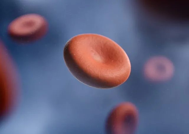
Iron is essential for oxygen transport and energy production. However, iron must remain tightly bound to proteins. Free iron can react with oxygen to form highly reactive molecules.
This process can damage cell membranes and trigger inflammatory signals. Poor iron regulation has been linked to oxidative stress and tissue injury (Galaris et al., 2019).
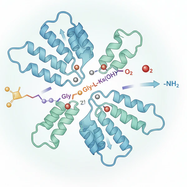
Copper & Iron Control
Mineral balance plays a key role in how the body manages iron and inflammation. Ceruloplasmin is a protein made in the liver that depends on copper. It helps convert iron into a form that can move safely through the blood.
This process allows cells to release stored iron without leaving reactive iron inside tissues (Harris et al., 1999).
Iron Movement In The Body
Iron normally travels attached to carrier proteins. These systems prevent free iron from accumulating in cells.
When copper dependent enzymes are impaired, iron may remain trapped inside tissues. This buildup can increase oxidative stress and stimulate inflammatory pathways (Collins, 2021).
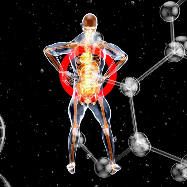
Effects Of Long Term Inflammation
Persistent inflammation can slowly damage tissues and organs throughout the body. Inflammatory signals can affect the lining of blood vessels. Over time, this may contribute to plaque formation and vascular injury.
Iron driven oxidative reactions are thought to play a role in these processes within the vessel wall (Cornelissen et al., 2019).
Brain & Nerve Stress
Iron balance is also important in the brain. When iron accumulates in neural tissue, oxidative stress may increase.
This stress has been linked to neurodegenerative changes and inflammation within the nervous system (Ward et al., 2014).
Metabolic Disruption
Chronic inflammation can interfere with normal metabolic signaling. This can affect how the body manages energy, blood sugar, and liver function. Inflammation and metabolic dysfunction often reinforce each other, creating a cycle that maintains tissue stress (Hotamisligil, 2006).
Consult a licensed healthcare professional before starting, stopping, or changing any diet, supplement, medication, or wellness practice. For questions about a medical condition or symptoms, seek advice from a qualified clinician who can assess your situation.
FAQs
What is the main purpose of inflammation?
Inflammation protects the body from injury and infection. It helps remove damaged cells and begin tissue repair.
Why can inflammation become harmful?
When inflammatory signals remain active for long periods, they can damage tissue and disrupt normal organ function.
How does iron influence inflammation?
Iron can generate reactive molecules when not properly controlled. These molecules can damage cells and trigger inflammatory responses.
What role does copper play in iron metabolism?
Copper supports enzymes such as ceruloplasmin that allow iron to move safely through the body and out of tissues.
Can inflammation affect many organs at once?
Yes. Chronic inflammation can influence blood vessels, the brain, the liver, and metabolic tissues.
Related Posts

Research
Collins, J.F., 2021. Copper nutrition and biochemistry and human (patho)physiology. Advances in Food and Nutrition Research.
Cornelissen, A., Guo, L., Sakamoto, A., Virmani, R. and Finn, A.V., 2019. New insights into the role of iron in inflammation and atherosclerosis. EBioMedicine.
Furman, D. et al., 2019. Chronic inflammation in the etiology of disease across the life span. Nature Medicine.
Galaris, D., Barbouti, A. and Pantopoulos, K., 2019. Iron homeostasis and oxidative stress: An intimate relationship. Biochimica et Biophysica Acta – Molecular Cell Research.
Gutteridge, J.M.C. and Halliwell, B., 2018. Oxidative stress, redox stress or redox success? Biochemical and Biophysical Research Communications.
Harris, Z.L., Durley, A.P., Man, T.K. and Gitlin, J.D., 1999. Targeted gene disruption reveals an essential role for ceruloplasmin in cellular iron efflux. Proceedings of the National Academy of Sciences.
Hotamisligil, G.S., 2006. Inflammation and metabolic disorders. Nature.
Medzhitov, R., 2008. Origin and physiological roles of inflammation. Nature.
Pradhan, A.D. et al., 2001. C-reactive protein, interleukin 6, and risk of developing type 2 diabetes mellitus. Journal of the American Medical Association. DOI: 10.1001/jama.286.3.327. PMID: 11466099
Visser, M. et al., 1999. Elevated C-reactive protein levels in overweight and obese adults. Journal of the American Medical Association. DOI: 10.1001/jama.282.22.2131. PMID: 10591334
Bo, S., Durazzo, M., Gambino, R., Berutti, C., Milanesio, N., Caropreso, A., Gentile, L., Cassader, M., Cavallo-Perin, P. and Pagano, G., 2008. Associations of Dietary and Serum Copper with Inflammation, Oxidative Stress, and Metabolic Variables in Adults ,. The Journal of Nutrition, [online] 138(2), pp.305–310. https://doi.org/10.1093/jn/138.2.305.
Batey RG, Lai Chung Fong P, Shamir S, Sherlock S. A non-transferrin-bound serum iron in idiopathic hemochromatosis. Dig Dis Sci. 1980 May;25(5):340-6. doi: 10.1007/BF01308057. PMID: 7371472.
Thackeray EW, Sanderson SO, Fox JC, Kumar N. Hepatic iron overload or cirrhosis may occur in acquired copper deficiency and is likely mediated by hypoceruloplasminemia. J Clin Gastroenterol. 2011 Feb;45(2):153-8. doi: 10.1097/MCG.0b013e3181dc25f7. PMID: 20502350.
Libby, P., Ridker, P.M. and Maseri, A., 2002. Inflammation and atherosclerosis. Nature. DOI: 10.1038/nature01323. PMID: 11961147
Prohaska, J. R. (2011). Impact of Copper Limitation on Expression and Function of Multicopper Oxidases (Ferroxidases). Advances in Nutrition, 2(2), 89-95. https://doi.org/10.3945/an.110.000208
Sorenson, J.R.J., 1989. 6 Copper Complexes Offer a Physiological Approach to Treatment of Chronic Diseases. Progress in Medicinal Chemistry, [online] pp.437–568. https://doi.org/10.1016/s0079-6468(08)70246-7.
Uriu-Adams, J.Y. and Keen, C.L., 2005. Copper, oxidative stress, and human health. Molecular Aspects of Medicine, [online] 26(4–5), pp.268–298. https://doi.org/10.1016/j.mam.2005.07.015.
Vashchenko, G., & A. MacGillivray, R. T. (2013). Multi-Copper Oxidases and Human Iron Metabolism. Nutrients, 5(7), 2289-2313. https://doi.org/10.3390/nu5072289
Wang, J. and Pantopoulos, K., 2011. Regulation of cellular iron metabolism. Biochemical Journal, [online] 434(3), pp.365–381. https://doi.org/10.1042/bj20101825.
Wallander, M.L., Leibold, E.A. and Eisenstein, R.S., 2006. Molecular control of vertebrate iron homeostasis by iron regulatory proteins. Biochimica et Biophysica Acta (BBA) – Molecular Cell Research, [online] 1763(7), pp.668–689. https://doi.org/10.1016/j.bbamcr.2006.05.004.
Xia, Y., Chen, Z., Huang, C., Shi, L., Ma, W., Chen, X., Liu, Y., Wang, Y., Cai, C., Huang, Y., Liu, W., Shi, R. and Luo, Q., 2024. Investigation the mechanism of iron overload-induced colonic inflammation following ferric citrate exposure. Ecotoxicology and Environmental Safety, [online] 275, p.116241. https://doi.org/10.1016/j.ecoenv.2024.116241.