Key Takeaways
- Ferroptosis begins when iron drives fast damage in fragile fats within cell membranes.
- Seed oil fats raise the pool of membrane fats that oxidize with less stress.
- Low glutathione and weak GPX4 leave cells less able to clear lipid damage.
- Iron overload gives cells more reactive iron that can push oxidation forward faster.
- The main danger comes when oxidized membrane lipids rise faster than repair systems.
What Ferroptosis Means
The Core Event
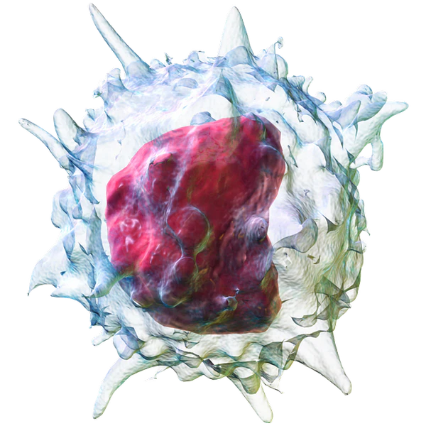
Ferroptosis is a form of cell death driven by iron and oxidized fats in the cell membrane. The membrane is the soft outer layer that helps keep the cell whole and working.
When too many membrane fats become oxidized, the membrane loses strength and the cell starts to fail.
Review papers in this field now describe that iron driven phospholipid damage as the main event, not a side detail of general stress (Jiang et al., 2021).
Polyunsaturated fats have more weak spots, so they are easier to damage during oxidation. In ferroptosis research, the most important target is not just any fat.
It is polyunsaturated fat built into membrane phospholipids, because those fats can become lipid peroxides, which means oxidized fats that injure the membrane from within (Dixon and Olzmann, 2024).
The process builds as oxidized membrane lipids rise faster than the cell can clear them.
Once that balance breaks, damage spreads through the membrane and the cell crosses a point it cannot reverse (Stockwell, 2022).
Why Iron Drives It
Cells need iron for energy, oxygen use, and many normal tasks. Trouble starts when too much iron is free inside the cell rather than bound and controlled.
That loose iron helps fuel reactions that turn vulnerable fats into damaged fats much faster.
Researchers often focus on ferrous iron because it can help generate highly reactive radicals. Those radicals attack membrane fats and help start chain reactions of lipid damage.
When iron handling goes off course, ferroptosis becomes much easier to trigger, especially in tissue already under stress (Ru et al., 2024).
The more reactive iron a cell carries, the less room it has before fat damage starts to run ahead of repair.
Why Seed Oils Matter
Fragile Fats
Seed oils are rich in linoleic acid, which is a polyunsaturated fat. In the body, polyunsaturated fats can become part of cell membranes.
Ferroptosis research shows that membranes loaded with these more fragile fats are easier to oxidize than membranes built from more stable fats.
This is one reason the field keeps coming back to membrane lipid makeup as a key part of ferroptosis sensitivity (Jiang et al., 2021).
Enzymes such as ACSL4 help place these vulnerable fats into membrane phospholipids. When more of these fats are placed into the membrane, the cell becomes easier to push into ferroptosis.
Iron provides the spark and fragile membrane fats provide the material that burns (Dixon and Olzmann, 2024).
Ferroptosis depends on oxidation prone membrane fats, and seed oils are a major source of those fats in modern diets.
The fats that make ferroptosis possible are the same kind of fats that seed oils supply in large amounts.
What Diet Can Show
Most ferroptosis papers study cells, animals, or disease models rather than long human feeding trials.
Ferroptosis requires vulnerable polyunsaturated membrane lipids, and seed oils increase intake of those same kinds of fats.
A membrane built with more oxidation prone fats is easier to damage than a membrane built with more stable fats. It does mean careful wording is needed.
The research gives strong mechanistic support for concern about seed oils in ferroptosis, but it does not yet give neat real world trial answers for every disease and every person (Stockwell, 2022).
Ferritin Is Not A Full Iron Answer
Ferritin can rise with inflammation, not only stored iron.
Defense Systems
Glutathione & GPX4
Cells have a cleanup system meant to stop this damage before it spreads. Two major parts are glutathione and GPX4.
Glutathione is a small antioxidant the cell uses to control oxidative damage. GPX4 is an enzyme that uses glutathione to reduce lipid peroxides, which means it helps neutralize oxidized membrane fats before they tear through the membrane.
When cystine import falls, glutathione drops. When glutathione drops, GPX4 cannot do its job well. When GPX4 is blocked or lost, lipid peroxides build up fast.
That leaves the membrane exposed and makes ferroptosis much easier to trigger (Zhang et al., 2024).
This is why ferroptosis is often described as a balance problem. The cell may handle some oxidative stress for a time, but once peroxide cleanup falls behind, membrane injury can spread very quickly.
Backup Defenses
GPX4 is not the only defense. FSP1 is another known system that can slow ferroptosis through a different route. It helps protect membrane lipids even when the glutathione system is under strain.
When that backup system is weak, cells lose another layer of protection and become easier to push toward ferroptosis (Dai et al., 2024).
Ferroptosis is not caused by iron alone and not caused by weak antioxidant defense alone.
The problem grows when both sides move in the wrong direction at once. Iron rises, fragile fats are present, and the defense systems fail to keep up.
Check vs Skip
| Check | Skip |
|---|---|
| Ferritin | Iron supplements |
| Transferrin saturation | Ferritin alone |
| Hemoglobin | Ignoring inflammation |
| Ceruloplasmin | Ignoring copper |
Common Triggers
Iron Overload
Iron overload is one of the clearest upstream triggers. This can happen through excess iron intake, repeated transfusions, inherited disorders of iron handling, or tissue level iron buildup during disease.
Once free iron increases inside cells, radical chemistry becomes easier and membrane fats become harder to protect (Ru et al., 2024).
Cells do try to store iron safely in ferritin and move it with control. Problems begin when storage, transport, or release goes off course.
A larger reactive iron pool gives ferroptosis a stronger push even before obvious symptoms appear.
Stress & Injury
Other triggers can push cells closer to the same endpoint. Ischemia reperfusion injury is one example.
Tissue first loses blood flow and then gets it back, which can create a burst of oxidative stress.
Some toxins, some cancer drugs, mitochondrial stress, inflammation, and glutathione depletion can also move a cell toward ferroptosis when the conditions are right (Patanè et al., 2023).
These triggers do not define ferroptosis by themselves. They become important because they add pressure to the same weak spots.
They raise oxidative stress, disturb iron handling, drain glutathione, or increase membrane lipid damage.
Copper & Ceruloplasmin
Copper helps the body handle iron with tighter control because ceruloplasmin needs copper to work well.
Ceruloplasmin has ferroxidase activity, which helps shift iron into a form that can move more safely in blood and tissues.
Human feeding data are direct on this point. In a controlled study, drinks providing twenty five percent of energy as glucose or fructose lowered ceruloplasmin ferroxidase activity, and glucose also lowered serum copper.
Ceruloplasmin protein did not change during the study, so the drop was in function rather than protein amount (Harder et al., 2020).
You can therefore have ceruloplasmin present while having weaker copper linked iron handling.
Lower copper status and lower ceruloplasmin activity can leave iron less controlled inside tissues, which can add to oxidative stress and support iron driven lipid damage.
Carbohydrates & Iron Handling
High carbohydrate intake, especially from sugar sweetened drinks, can weaken iron handling through effects on copper and ceruloplasmin.
The same controlled feeding study found that glucose and fructose lowered ceruloplasmin ferroxidase activity within a short period, while glucose also lowered serum copper (Harder et al., 2020).
High glucose can also interfere with vitamin C handling during hyperglycemia.
Glucose competes with transport of the oxidized form of vitamin C into some cells, which can weaken antioxidant protection where it is needed most (Chen et al., 2005) (Liu et al., 2022).
A high carbohydrate load can impair copper status, lower ceruloplasmin activity, and weaken part of the system that helps keep iron under control.
The Simple View
The direct cause of ferroptosis can be said in plain words. Iron drives oxidation of fragile membrane fats faster than the cell can stop it. Seed oils fit the story because they supply the kind of fats that make membranes easier to oxidize. Iron overload fits the story because it raises the reactive iron that pushes the damage forward.
For any health concerns or questions about a medical condition, get guidance from a physician or another appropriately trained clinician. Before changing your diet, supplements, or health routine, talk with a licensed healthcare professional.
Suggested Posts

Evidence Limits
Research
Jiang, X., Stockwell, B.R. and Conrad, M. (2021) ‘Ferroptosis: mechanisms, biology and role in disease’. Nature Reviews Molecular Cell Biology, 22(4), pp. 266–282.
Dixon, S.J. and Olzmann, J.A. (2024) ‘The cell biology of ferroptosis’. Nature Reviews Molecular Cell Biology, 25(6), pp. 424–442.
Stockwell, B.R. (2022) ‘Ferroptosis turns 10: Emerging mechanisms, physiological functions, and therapeutic applications’. Cell, 185(14), pp. 2401–2421.
Ru, Q., Wen, S., Li, W., Huang, J., Chai, D., Cao, Y., Lin, W., Yao, Y., Li, Y. and Tang, D. (2024) ‘Iron homeostasis and ferroptosis in human diseases: mechanisms and therapeutic prospects’. Signal Transduction and Targeted Therapy, 9, 271.
Zhang, W., Liu, Y., Wang, T. and Yao, Y. (2024) ‘GPX4, ferroptosis, and diseases’. Biomedicine and Pharmacotherapy, 173, 116493.
Dai, Q., Ma, J., Zhu, C., Zuo, M., Li, X., Liu, X. and Li, H. (2024) ‘Inhibition of FSP1: A new strategy for the treatment of tumors’. Oncology Reports, 52(2), 133.
Patanè, G.T., Fazio, G., Siliberto, G., Rapisarda, V. and Ledda, C. (2023) ‘Ferroptosis: Emerging role in diseases and potential implication of bioactive compounds’. International Journal of Molecular Sciences, 24(24), 17279.
Berndt, C., Friedmann Angeli, J.P., Conrad, M., Dixon, S.J. and Olzmann, J.A. (2024) ‘Ferroptosis in health and disease’. Redox Biology, 75, 103211.
Tang, D., Chen, X., Kang, R. and Kroemer, G. (2021) ‘Ferroptosis: molecular mechanisms and health implications’. Cell Research, 31(2), pp. 107–125.
Yan, H.F., Zou, T., Tuo, Q.Z., Xu, S., Li, H., Belaidi, A.A. and Lei, P. (2021) ‘Ferroptosis: mechanisms and links with diseases’. Signal Transduction and Targeted Therapy, 6(1), 49.
Stockwell, B.R., Friedmann Angeli, J.P., Bayir, H., Bush, A.I., Conrad, M., Dixon, S.J., Fulda, S., Gascón, S., Hatzios, S.K., Kagan, V.E., Noel, K., Jiang, X., Linkermann, A., Murphy, M.E., Overholtzer, M., Oyagi, A., Pagnussat, G.C., Park, J., Ran, Q., Rosenfeld, C.S., Salnikow, K., Tang, D., Torti, F.M., Torti, S.V., Toyokuni, S., Woerpel, K.A. and Zhang, D.D. (2017) ‘Ferroptosis: A regulated cell death nexus linking metabolism, redox biology, and disease’. Cell, 171(2), pp. 273–285.
Zou, Y. and Schreiber, S.L. (2020) ‘Progress in understanding ferroptosis and challenges in its targeting for therapeutic benefit’. Cell Chemical Biology, 27(4), pp. 463–471.
Tang, D. and Kroemer, G. (2020) ‘Ferroptosis’. Current Biology, 30(21), pp. R1292–R1297.
Seibt, T.M., Proneth, B. and Conrad, M. (2019) ‘Role of GPX4 in ferroptosis and its pharmacological implication’. Free Radical Biology and Medicine, 133, pp. 144–152.
Xie, Y., Hou, W., Song, X., Yu, Y., Huang, J., Sun, X., Kang, R. and Tang, D. (2016) ‘Ferroptosis: process and function’. Cell Death and Differentiation, 23(3), pp. 369–379.
Cao, J.Y. and Dixon, S.J. (2016) ‘Mechanisms of ferroptosis’. Cellular and Molecular Life Sciences, 73(11–12), pp. 2195–2209.
Zhong, M., Lei, P., Anderson, G.J. and Wang, W. (2025) ‘Iron metabolism and ferroptosis in human health and disease’. Biology Direct, 20, 83.
Li, S., Yang, X., Hao, Y., Wang, M., Gao, Y., Chen, W. and Lin, M. (2024) ‘Ferroptosis is a protective factor for the prognosis of cancer patients: a systematic review and meta analysis’. BMC Cancer, 24, 604.
Coradduzza, D., Congiu, T., Schepis, T., Beretta, G.L. and Tinelli, A. (2023) ‘Ferroptosis and senescence: A systematic review’. International Journal of Molecular Sciences, 24(4), 3658.
Zhao, J., Wang, H., Huang, Y., Zhang, Y., Liu, Y., Li, Y., Qiao, Y., Cui, L., Song, M., Zhang, C., Wang, J., Wang, Z., Liu, X., Wang, Y., Caceres, C., Tani, H., Wu, Y., Guo, F., Smith, A., Liu, J., Liu, H., Yu, J., Sun, Y. and Cheng, T. (2023) ‘Human hematopoietic stem cell vulnerability to ferroptosis’. Cell, 186(4), pp. 732–747.e16.